CLOSE

Hematological tumors

Hematologic malignancies, also called blood cancers are tumors that affect cells of the bone marrow, the lymphatic and the immune system. Basically, hematological malignancies originate from the proliferation and the survival of the two major blood cell lineages: myeloid and lymphoid cell lines.

Select your topic of interest

  1.  Leukemia (cancer of the blood cells)  

  1. Leukemia (cancer of the blood cells) can be of four types: 
    1. Acute myeloid leukemia (AML)
    2. Acute lymphoblastic leukemia (ALL)
    3. Chronic myelogenous leukemia (CML)

Chronic lymphocytic leukemia (CLL)


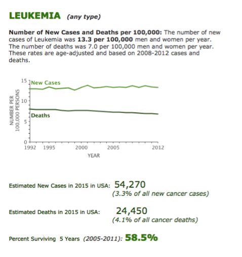
Figure 5.The most relevant epidemiological data on Leukemias, reported by the USA-SEER Agency. 


Acute Leukemia, Myeloblastic (AML) or Lymphoblastic (ALL)

Acute leukemia is a type of fast growing blood cancer that requires prompt treatment. Blood is made up of different types of cells that are produced in the interior of bones in a part called the bone marrow. When people have leukemia, their bone marrow produces abnormal blood cells that are out of control and are not effectively working according to the body needs. This can cause signs and symptoms such as

  • Feeling fatigues and weak
  • Bleeding
  • Fever and easily catching infections


The diagnosis:

Successful therapeutic plan of all blood cancers depends on the proper diagnosis at the microscopic and molecular level. This is done through comprehensive evaluation of:

  • Blood tests, in particular Complete Blood Count (CBC) and blood film examination
  • Bone marrow examinations, including:
    • Histological evaluation
    • Immunophenotypic analysis of blood and marrow cells
    • Cytogenetic analysis to detect chromosomal abnormalities
    • Genetic studies in order to detect disease-specific DNA abnormalities


Treatment for acute leukemia:

Treatment is basically complex and depends on the type of leukemia and other factors such as age and donor availability for bone marrow transplant.

Treatment of acute leukemia generally includes two main initial parts.

  • The first part of treatment is called “induction of remission” and is approximately 4 weeks. This part of treatment requires you to stay in the hospital and receive chemotherapy regimens.
    • Once in “remission” state, which means no abnormal blood cells are detected in the blood or bone marrow by microscopic examination, the second part starts, the so-called “post-remission therapy.” This part includes:

additional chemotherapy courses, for 3 to 4 months period and, in younger patients,

bone marrow transplant: either by using the patient own bone marrow cells to support the use of higher doses of chemotherapy (autologous transplant) or replacing the diseased bone marrow cells with more heathy cells from a volunteer donor (allogeneic transplant or allo-HSCT, i.e. allogeneic-Hematopoietic Stem Cell Transplantation). In fact, alloHSCT represents the most effective option for patients younger than 65-70 yrs., with Acute Leukemia and unfavorable prognostic features or with refractory or relapsed disease; the immune-mediated anti-leukemia effect, which is peculiar of allogeneic transplantation, makes the allo-HSCT procedure extremely effective in Leukemia although much more complex compared to autologous transplantation. 

ü  Radiation therapy is sometimes used to kill remaining leukemia cells at certain organs of the body like in cases of central nervous system involvement, usually at the end of chemotherapy courses.


The importance of disease assessment following therapy

  • The accurate evaluation of response to therapy is crucial in Acute Leukemia (AL). In particular, molecular and immunophentypical procedures have been developed in order to identify minimal quantities of tumor cells that might persist following treatment below the levels detectable by standard methods of evaluation. This is called Minimal Residual Disease (MRD). The evaluation of MRD has reached a key role in the management of AL, as well as in other hematological malignancies where MRD can be assessed and monitored. In particular, in AL MRD may direct the treatment strategies following induction therapy and the preferred use of additional chemotherapy versus transplant-based is based quite often on results of MRD. Also at our Institutions at IEO, the study of MRD both in bone marrow (BM) and peripheral blood (PB) cells is a key point in the management of AL patients. In addition, research studies are ongoing with a dual aim:  to optimize the use of MRD analysis in AL patients and to extend the population of patients with hematological malignancies that might be assessed and monitored by MRD.         
  • MRD is regularly assessed at given time points during treatment. Moreover, all patients undergo regular visits, with blood tests and additional clinical exams, if needed, during the various treatment steps and at long term once the whole treatment program is completed


Within our OM-2 Division at IEO, there are sub-unit of doctors with specific interest in each of the main hematological malignancies, i.e. Acute Leukemia with the alloHSCT program, Lymphoma and Multiple Myeloma. The doctors mainly involved in the management of Acute Leukemia and in the allogeneic transplant procedures are: Dr. Rocco Pastano (Director of the allogeneic-HSCT program), Dr. Federica Gigli and Dr. Simona Sammassimo along with the support of Dr. Safaa Ramadan   

Chronic leukemia

 Chronic Lymphocytic Leukemia (CLL), and Chronic Myeloid Leukemia (CML) 

Chronic leukemia is a type of blood cancer that usually grows slowly.

Main signs and symptoms are:

  • Feeling fatigues and weak
  • Easily catching infections
  • Fevers, drenching sweats at night, and losing weight
  • Lymphadenopathy and organomegally: patients with Chronic Lymphocytic Leukemia (CLL) may have swollen lymph nodes in the neck, under the arm, or in the groin; patients with Chronic Myeloid Leukemia (CML) may present with large spleen.


The diagnosis:

  • CBC (Complete Blood Count), along with blood film analysis, is the first step to suspect a Chronic Leukemia
  • The diagnosis of CLL can be made by Immunophenotypic Analysis, that may show the presence of the a clonal Lymphocyte population, i.e. the neoplastic CLL population
  • CML diagnosis requires the detection by molecular analysis of blood or marrow DNA, revealing the presence of the distinctive bcr-abl gene, that originate by the translocation involving chromosomes 9 and 22, giving rise to the diagnostic “Philadelphia Chromosome” (Ph-1)  


Treatment of chronic leukemia:

Chronic lymphocytic leukemia (CLL):

Being slowly growing and indolent tumor, CLL is treated when the disease causes symptoms or starts growing faster than its original rate.

The main form of treatment is chemotherapy combined with immunotherapy. The choice between different chemotherapy drugs is based not only on the molecular profile of the disease but also on patient related factors and comorbidities. Immunotherapy is mainly represented by monoclonal antibodies directed against surface antigens of neoplastic Lymphocyte. By far, the most extensively employed is the anti-CD20 monoclonal antibody Rituximab. Other monoclonal antibodies and novel drugs directed against molecular checkpoints that are abnormally expressed in CLL cells are under advanced development (see also: Lymphoma section, Novel targeted drugs)


Chronic myeloid leukemia (CML):

CML cells harbor the abnormal bcr-abl gene, whose product is a tyrosine-kinase responsible for the over-production of myeloid blood cells. In the last two decades, very effective drugs inhibiting the bcr-abl tyrosine-kinase product (known as Tyrosine-kinase inhibitors or TKIs) have been developed, with a of great consequence for the management and the long-term life-expectancy for patients with CML. Nowadays, most CML patients are successfully treated with TKIs, without need of neither chemotherapy nor transplant procedures . TKIs are very effective in long term control of the disease and resulted in a revolutionary success in the history of this disease. Treatment requires frequent visits to evaluate the course of the disease response with frequent blood and as necessary bone marrow exams.

In few cases, the cancer blood cells grows faster and becomes less controlled by the “first generation” TKI; however, newly developed, “second or third generation” TKIs, may overcome drug-resistance.


Other treatment options, which can help to control the usually infrequent advanced stages of the disease are:

  • Chemotherapy: a number of regimens can be available.
  • Bone marrow transplant: after intensive chemotherapy, a healthy donor bone marrow cells are used to replaces cells in the bone marrow of the patient.


Staff

The doctors mainly involved in the management of Acute Leukemia and in the allogeneic transplant procedures are: Dr. Rocco Pastano (Director of the allogeneic-HSCT program), Dr. Federica Gigli and Dr. Simona Sammassimo along with the support of Dr. Safaa Ramadan   


  1.  Lymphoma  (cancer of the lymphatic cells) 

Lymphoma is a cancer that affects the lymphatic system organs. The lymphatic system is an important part of the immune system. When lymphoma cells grow out of control, they result in swelling of the lymph nodes and can spread at different parts of the body.

There are two main types of lymphoma known as

  • > Non-Hodgkin’s Lymphoma (NHL): there are several subtypes, the most frequent are diffuse large B-cell lymphoma (DLB-CL) and follicular lymphoma (FL).
    • > Hodgkin Lymphoma (HL)

Non-Hodgkin’s Lymphoma is by far more common than Hodgkin’s Lymphoma. Overall, Lymphoma represent the most frequent type of hematological malignancy, as shown in Figure 1.


Details, of the most relevant epidemiological data on non-Hodgkin’s and Hodgkin’s Lymphoma, reported by the USA-SEER Agency, are shown in Figure 6 and 7. 


 Presentation and symptoms:

The most common presentation of the disease is swollen lymph node in one or more sites of the body, such as in the neck, armpit, groin, as well as in the thorax and abdomen. Patients may also have fever, weight loss or wake up at night with remarkable sweats that soak their cloths.


Main tests for diagnosis:

  • Lymph node biopsy and histological diagnosis: excision of the swollen lymph node for miscrospic examination is the most critical step in the diagnosis. 
  • Due to the multiple subtypes of NHL and HL, the histological examination is fundamental for the appropriate management and treatment of all patients with a lymphoproliferative disease. In this view, the IEO has instituted a dedicated Unit for the histological diagnosis of hematological neoplasms, with particular focus on Lymphoma. The Unit is directed by Prof. Stefano Pileri, which is a highly renowned expert in the field of pathological diagnosis of lymphoproliferative diseases. Indeed, due to his high reputation, Prof. Pileri is one of the eight Editors of the 2008 WHO Classification of Tumors of Hematopoietic and Lymphoid Tissues (see Figure on the right), which is now ready for the 2016 update.
  • Imaging techniques: CT scan, PET scan and ultra-songraphy are the common imaging procedures employed to evaluate the extent of the disease in the body (staging procedure)
  • Bone marrow biopsy is usually needed in order to investigate possible bone marrow involvement by the disease


Treatments for lymphoma:

The treatment plan is quite variable, depending on several factors, namely: i. histological type/subtype of the lymphoma; ii. extent of disease dissemination; iii. patient age; iv.  patient characteristics and comorbidities. In general, slowly growing forms with no symptoms may not need any treatment or mild treatment schedules, while fast growing lymphomas necessitate immediate and often intensive therapy.


The main treatment options currently available are here summarized:

  • Chemotherapy: a number of regimens, i.e. combinations of chemotherapeutic agents, have been developed and validated for the treatment of the fast growing lymphomas. These regimens are regularly administered in repeated courses and they still represent the main treatment options for several lymphoid neoplasms.
  • Immunotherapy with humanized Monoclonal Antibody: Humanized Monoclonal antibodies have been introduced in the clinical practice during the last years of 1900. This has produced an unprecedented advancement in the management of most lymphoid neoplasms. By far, the most representative and highly effective is the anti-CD20 Rituximab Monoclonal Antibodies, which has been widely employed, either alone or in combination with chemotherapy, over the last 15 years in the management of virtually all B-cell lymphoma, including the most frequent DLBCL and FL forms. The availability of Rituximab has resulted in significant improvements in cure rate and in the overall life expectancy for most B-cell lymphoma. Several novel antibodies targeting various tumor-associated antigens have been developed over the last 15 years and studies are still ongoing aimed to optimize the therapeutic use of monoclonal antibodies and other immunotherapeutic drugs.
  • Radiation therapy: it represent an additional treatment option in the management of lymphoma. It is usually used to kill remaining lymphoma cells at end of chemotherapy courses or as alternative option in case of disease recurrence and reduced tolerability to systemic treatments.
  • Bone marrow transplantation: this type of transplant is done using the same person bone marrow cells. The bone marrow cells that generate the blood elements are collected. The patient then receives a more intensive chemotherapy regimen that aims to kill all cancer cells but it also kill the bone marrow cells. So, after the intensive chemotherapy, the collected bone marrow cells are put back in the patient body to support him during the period needed for recovery from the chemotherapy effect
  • Novel targeted drugs: In the last decade, impressive advancements have been obtained in the understanding of normal and pathological lymphocyte generation, with major contributions by some eminent research groups, including the group directed by Riccardo Dalla Favera at the Columbia University in New York. This has lead to the identification of several molecular checkpoints that are crucial for the proliferation of lymphoma cells. As a consequence, novel lymphoma subtypes have been recognized and the WHO Classification is the unique reference text (see the front-page reported in Figure 8) for the correct pathological diagnosis of any lymphoma entity that can be now identified on the basis of specific cellular and molecular markers. As mentioned, Prof. Stefano Pileri, Director of the Hematophatology Unit at IEO, is among the eight Editors of the WHO Classification. The recently identified molecular markers have been viewed as potential therapeutic targets. Thus, novel drugs have been developed and others are under development to target molecular checkpoints that might be specifically altered in each lymphoma subtype. The present challenge is to define the ideal place of these new drugs, in combination with other effective treatment already available for lymphoma. The accurate and detailed histological diagnosis for any new patient is now mandatory to optimally exploit the novel molecularly-directed drugs.


Staff

Within the OM-2 Division, the doctors mainly involved in the management of Lymphoma (including Hodgkin’s and non-Hodgkin’sLymphoma and CLL) are: Dr. Alberto De Crescenzo, Dr. Anna Vanazzi, Dr. Angela Gueli and Dr. Enrico Derenzini, along with the support of Dr. Safaa Ramadan

  1. Multiple myeloma  (cancer of plasma cells) 


The most relevant epidemiological data on Myeloma, reported by the USA-SEER Agency, are shown in Figure 9.


Multiple myeloma (MM) is a cancer that occur in a type of white blood cells called plasma cells (PC).

Plasma cells are important to the body’s immune system. Indeed, PC are committed to the production of Immunoglobulins, i.e. our antibodies. In dedicated deputy In MM, there is the massive expansion of a clonal population of PC that usually produces a single specific type of Immunoglobulin or part (light chains) of a given Immunoglobulin.

The bone marrow is markedly affected by the expansion of abnormal PC, with several consequences, including an inefficient immune activity.

Presentation, symptoms and diagnosis

  • Main symptoms are:

Bone pain

Weakness, easy fatigue

Nausea, vomiting, confusion, or feeling more thirsty

 Numbness, tingling, or weakness

Losing weight without changing of eating habits


  • Main tests for diagnosis:

 Blood and urine tests

 Bone marrow biopsy

 Imaging: X-ray or MRI and others, with particular attention to the study of bones

 
The most relevant epidemiological data on Myeloma, reported by the USA-SEER Agency, are shown in  the figure above.


Treatment of Multiple Myeloma:

Multiple myeloma can follow a very slow course and causes no manifestations (smoldering myeloma) or present with more rapidly growing diseases and distressing symptoms. So treatment decision varies according to many factors including the disease nature and the patient general condition and comorbidities.

In general treatment options can be:

  •  
  • > Observation and follow up without any specific therapy, for patients with smoldering form of MM
  • > Active treatment, for all patients presenting with more active disease, includes: 
    • Chemotherapy: a number of protocols of chemotherapy drugs are available in the treatment of multiple myeloma. The selection is based on                patient and disease related feature and the specificity and toxicity profile of each drug.
    • Steroid medicines: are important part in the treatment that potentiate the killing of cancer cells
    • A class of drugs known as immune modulating, have been introduced in the treatment of MM, over the last 15 years. These drugs can arrest              the growth of cancer cells and are usually employed either alone or most often in combination treatments
    • BM transplant: this type of transplant is done using the same person bone marrow cells and the procedure is known as auto-transplantation. The bone marrow cells that generate the blood elements are collected. The patient then receives a more intensive chemotherapy regimen that aims to kill all cancer cells but it also kill normal BM cells. So, after the intensive chemotherapy, the collected BM cells are put back in the patient body to support him during the period needed for recovery from the chemotherapy effect. At present, auto-transplant is a major treatment option in MM, at least for patients aged <65 yrs.


Several treatment options are now available in MM; at present, the common approach includes the alternation of different treatment schedules, with the alternating delivery of steroids, immune-modulating drugs and conventional chemotherapy, either alone or in combination, and intensified chemotherapy with auto-transplant, as well, if feasible. Indeed, intensified chemotherapy with autotransplant is a key treatment option in MM, particularly in adult patients aged < 65 years. Programs extending the use of autotransplant-based therapy to elderly subjects are under evaluation   


The transplant procedure with hematopoietic stem cells from a compatible donor, i.e. the allogeneic transplantation (alloSCT), has a minor role in the management of MM. However, in younger patients, with reduced response to standard treatment, including the new immune-modulating drugs, the allogeneic transplantation may be an alternative and potentially effective treatment option


Staff

Within the OM-2 Division, the doctors mainly involved in the management of Multiple Myeloma are: Dr. Alberto Agazzi, Dr. Niccolò Frungillo, along with the support of Dr. Simona Sammassimo  


Myelodysplastic syndromes (serious blood abnormalities that can lead to overt cancer)

Myelodysplastic syndromes (MDS) are a group of conditions in which the bone marrow, at the centre of the bone, which produces the blood cells, does not work appropriately. It instead produces abnormal immature blood cells that are not able to function. In fact, hematopoietic function can be quite variably affect, with wide clinical features, ranging from mild anemic forms to severe BM impairment and clinical manifestations of a pre-leukemic phase. Indeed, there is now a defined classification of the various disease subtypes that are collectively included in the heterogeneous group of Myelodysplastic syndromes.


Main presentation and diagnosis

Disease presentation is quite variable, according to the different disease subtypes. Sometimes an initial form of MDS is recognized accidentally during a routine medical check or another indication, in other cases, clinical manifestation may be pronounced and rapidly worsening.


Symptoms could be:

  • Feeling fatigues and weak
  • Difficulty in breathing on effort
  • Bleeding easily
  • Easily catching infections


Main diagnostic tests are:

  • Blood tests
  • BM aspirate and BM biopsy: morphological examination of BM smear remain a key diagnostic tool, along with BM histological examination. In addition, several biologic investigations, including cytogenetic and molecular studies, are done on the taken samples to identify and characterize the type of MDS.


Treatment for Myelodysplastic Syndromes:

As expected, treatment varies widely, according to the type of MDS and the symptoms of the patient. So treatment can be one of the following:

  • Clinical monitoring and transfusion support, when needed: blood and platelets donated from volunteers can be given to support patients and relieve the symptoms related to anemia or bleeding tendency.
  • Medications: some drugs can variably induce the bone marrow to make more blood cells (hematopoietic cytokines and differentiating drugs), others are used to helping the immune system defense against microbes
  • Chemotherapy: when the disease becomes more aggressive, treatment with chemotherapy using regimens similar to those of acute leukemia is usually given to control the disease. New classes of chemotherapy are available to control the disease with less side effects than chemotherapy are also very effective treatment options.
  • Bone marrow transplant: after intensive chemotherapy, a healthy donor bone marrow cells are used to replaces the cells in the bone marrow of the patient. Allo-SCT remains a complex procedures, with morbidly and mortality risks. Usually, the procedure is considered only for younger patients, with specific clinical characteristics predicting an unfavorable outcome with non-transplant- based approaches


Staff

Within the OM-2 Division, the same Doctors who take care of Acute Leukemia patients are also in charge for patients with Myelodysplastic Syndrome

Other Hematological disorders 

Besides the most frequent hematological malignancies, that have been above reviewed, several other hematological disorders are managed by the team of specialist doctors and nurses of the Hemato-Oncology Division of the I.E.O. This is a short list, with some additional information, of the most representative hematological diseases not included in the previous sections:


Chronic Myeloproliferative Neoplasms: this is a group of diseases, characterized by the overproduction of blood cells by BM. The BM can make too many red blood cells, white blood cells, or platelets. Thus, chronic myeloproliferative neoplasms includes six different diseases, each characterized by abnormalities of specific hematopoietic cell lineage, that are here listed:


  • Chronic Myelogenous Leukemia (CML), that has been described above, under the Chronic Leukemia section
  • Polycythemia Vera (PV), characterized by overproduction or red blood cells
  • Primary Myelofibrosis (also called chronic idiopathic myelofibrosis), characterized by marrow fibrosis and expansion of the spleen, leading to abnormal splenomegaly
  • Essential thrombocythemia (ET), characterized by overproduction of platelets
  • Chronic neutrophilic leukemia, with overproduction of neutrophilic granulocyte, in the absence of the specific cytogenetic marker of CML
  • Chronic eosinophilic leukemia, with overproduction of eosinophilic granulocyte


 Main diagnostic tests are:

Blood tests with analysis of CBC (number of red blood cells, platelets, number and types of white blood cells), and morphological evaluation of PB smears, with particular attention to the shape of red blood cells and the presence of immature myeloid cells

BM aspirate and BM biopsy, with morphological examination of BM smear along with BM histological examination

additional biologic investigations on BM cells are now mandatory, including cytogenetic analysis  to look for certain changes in the chromosomes and molecular studies, with particular attention to identify possible mutations of the JAK-2 gene


Leukopenia (decrease in the number of white blood cells) - Anemia (decrease of hemoglobin and red blood cells) - Thrombocytopenia (decrease in the number of platelets): these are abnormalities that can be observed at routine laboratory controls and may be ascribed to a variety of underlying diseases. In some instances, these abnormalities of CBC can be due to an impaired BM function and appropriate diagnostic investigations should be planned, following hematologic consultation   

  • in distinct conditions, BM cellularity is markedly reduced, in other words BM is hypoplastic, with a variably low residual hematopoiesis. In the absence of any known reasons of marrow hypoplasia, the diagnosis can be of Aplastic anemia. BM function impairment causes a variable reduction of CBC, i.e. the so called “pan-cytopenia”. When CBC values are profoundly depressed, the diagnosis is of Severe Aplastic Anemia (SAA). Careful BM examination is mandatory whenever a condition of Aplastic Anemia is suspected. Once the diagnosis is confirmed, appropriate treatments should be promptly instituted. At present, there are two main treatment options for SAA: i. since SAA is often an immunological-driven disease, intensive immunosuppression can be delivered, along with BM stimulation by hematopoietic cytokines; ii. the alternative approach is to replace the impaired BM with normal hematopoietic stem cells donated by a volunteer donor, that is the alloSCT procedure 


 Monoclonal gammopaty of undetermined significance (MGUS):
The presence of an abnormal immunoglobulin (or monoclonal protein, or M protein) is usually termed as monoclonal gammopathy. Multiple myeloma (see section above) is the most typical malignant monoclonal gammopathy. However, there might be monoclonal gammopathy, in the absence of any clinical problem. This is the typical presentation of the Monoclonal Gammopathy of Undetermined Significance (MGUS), a frequently encountered, non-malignant condition. Indeed, the presence of a M-component at serum protein electrophoresis is not an uncommon finding. Usually, MGUS causes no problems. Sometimes this mild abnormality of the serum protein assay is associated with another disease, however the common feature of MGUS is its occurrence in healthy subjects, in the absence of any other associated disease. Additional serum and urine laboratory tests are usually performed, BM evaluation may be required in a few cases. The diagnosis of a suspected MGUS is made if the M-component does not increase at subsequent serum protein controls. In fact, patients with a suspected MGUS usually have periodic checkups to monitor the level of M protein. If there is no increase, monoclonal gammopathy does not require treatment. If MGUS does not display signs of progression, no treatments are required. In a minority of cases, the M-component may progressively increase over years to other disorders, including some forms of blood cancer. Thus, MGUS does not necessitate neither treatments nor prophylactic supports, however periodic clinical and laboratory controls are recommended, at 6-12 mos. intervals.


Disorders of the coagulation system:

The term “hemostasis” indicates the complex system that regulates the coagulation process of our body. Treatment of patients with abnormalities of hemostasis is an integral part of Hematology. Thus, also at the Hemato-Oncology Division of the I.E.O., there is a section specifically committed to the managing of patients presenting with thrombotic and/or hemorrhagic disorders. Dr. Alberto Agazzi is in charge for the hemostasis disorders, due to his long experience and knowledge in the field. The teamwork takes advantage of the assistance of Dr. Giacomo Tamponi, who is the external consultant. Thrombotic and hemorrhagic disorders are frequent complications of hematological malignancies, at presentation and during treatment. Moreover, disorders of the coagulation system may occur in non-hematological neoplasia as well as in the absence of any underlying malignancy. Overall, the hemostasis team of the Hemato-Oncology Division of the I.E.O. takes care and provide consultation for patients presenting with the various dysfunctions of the coagulation system


Useful Links

  • ARE YOU LOOKING FOR A DOCTOR?

    Search for a doctor by surname, organizational unit or the first letter of the surname.

  • DO YOU WANT TO BOOK AN APPOINTMENT?

    At IEO, it is possible to book specialist appointments, diagnostic tests and other outpatient services.

  • WOULD YOU LIKE TO JOIN AN EXPERIMENTAL PROGRAMME?

    Read the guidelines for participating in an trial programme

UN PICCOLO GESTO E' PER NOI UN GRANDE AIUTO

PARTNERSHIP

Università degli Studi di Milano

MAIN CREDITS

Ministero della Salute Joint Commission International bollinirosa

© 2013 Istituto Europeo di Oncologia - via Ripamonti 435 Milano - P.I. 08691440153 - Codice intermediario fatturazione elettronica: A4707H7

IRCCS - ISTITUTO DI RICOVERO E CURA A CARATTERE SCIENTIFICO